13
Cells in the making
- What do you see?
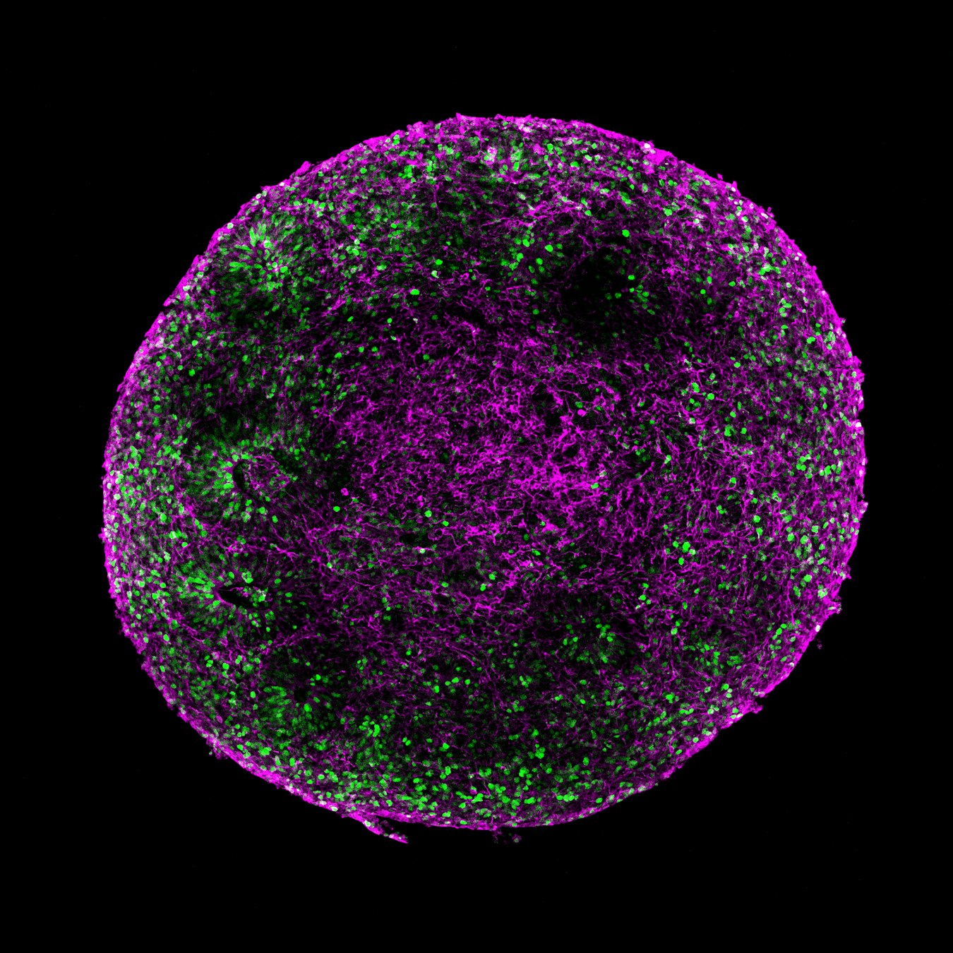
A cross-section of a human brain organoid, revealing cells at different stages of development. Some are young and still dividing, while others have matured into neurons and are beginning to form branching networks. - Green: Young cells that are still dividing
Pink: Mature neurons and their branching fibers
- Where is it found?
This miniature model of human brain tissue does not occur naturally in the body. Scientists grow it in the laboratory from human stem cells to recreate aspects of the developing midbrain.
- How big is it?
The organoid is approximately 2 millimeters in diameter.
≈ the size of a sesame seed
- What can we learn from this image?
Building brain tissue is a process of constant change. Some cells are still multiplying, while others have already begun to specialize and mature into neurons. Seeing both stages together allows scientists to follow how a miniature model of the brain takes shape over time.
- What are its superpowers?
Brain organoids can recreate important stages of human brain development in the laboratory. Their cells divide, specialize, mature, and begin to organize themselves, giving scientists a unique opportunity to observe processes that are extremely difficult to study in the developing human brain itself.
- How was this image taken?
This image was captured using confocal fluorescence microscopy. Fluorescent markers highlight cells at different stages of development, while the microscope reveals their organization within three-dimensional tissue.
- Image: Eurac Research / Ingrid Battistella – Neuroscientist
- What are scientists trying to understand with this image?
Scientists use these organoids to investigate how the human brain develops and how neurons grow and mature. They are particularly interested in dopamine-producing neurons like those that are gradually lost in Parkinson’s disease, with the goal of understanding what makes these cells vulnerable and how they might one day be protected.
The story behind the image
Inside this tiny piece of laboratory-grown brain tissue, development is happening at different speeds. Some cells are still dividing, creating building blocks for future tissue. Others have already become neurons and are extending delicate branches toward their neighbors. Captured together in a single image, they reveal brain development as a process in motion, with new cells continually emerging, changing, and finding their place.
Credits: Eurac Research / Ingrid Battistella