14
Neural bloom
- What do you see?
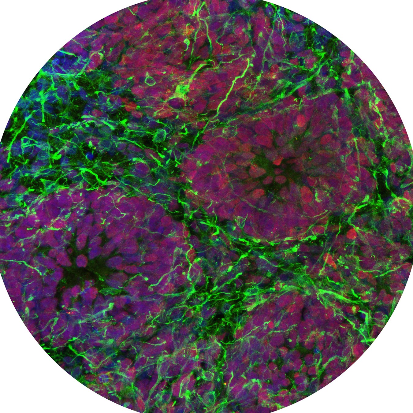
Flower-like structures called neural rosettes inside a human forebrain organoid. These circular arrangements of developing cells recreate some of the earliest architecture seen as the cerebral cortex begins to form. - Blue: Cell nuclei
Green: Neurons and their branching structures
Red: Neural progenitor cells, young cells that can develop into neurons of the cerebral cortex
- Where are they found?
These structures form inside laboratory-grown organoids made from human induced pluripotent stem cells. The organoids are designed to model the dorsal forebrain, the region that gives rise to much of the cerebral cortex.
- How big is it?
These organoids can grow to approximately 2 millimeters in diameter.
≈ the size of a sesame seed
- What can we learn from this image?
The developing brain is highly organized from its earliest stages. Young cells arrange themselves into characteristic patterns, creating structures from which neurons emerge and mature. By reproducing these patterns in organoids, scientists can observe some of the fundamental steps involved in building the human cerebral cortex.
- What are their superpowers?
Brain organoids can recreate important aspects of specific regions of the human brain in miniature. They allow scientists to observe how brain cells develop and organize, investigate what changes in neurological diseases, and explore how cells respond to potential new treatments.
- How was this image taken?
The organoid was frozen and cut into thin sections. The sections were mounted on glass slides and processed using immunofluorescence, a technique that labels specific cells and structures with fluorescent markers. The images were then captured using a confocal fluorescence microscope. - Image: Eurac Research / Sara Pizzi – Cellular Biologist
- What are scientists trying to understand with this image?
Scientists create organoids that represent different regions of the human brain and combine them into more complex models called assembloids. These models help researchers investigate how different brain regions develop, connect, and communicate, and what changes when neurological disorders occur.
The story behind the image
At first glance, these glowing structures could be flowers opening in the dark. In fact, they are groups of young brain cells arranging themselves into neural rosettes, one of the characteristic patterns seen during early brain development. These organized clusters are part of the construction process for the intricate architecture of the cerebral cortex. By recreating this process in the laboratory, scientists can see some of the earliest steps involved in building the human brain.
Links
Tiny blobs. Huge potential ⠀⠀⠀ Investigating Parkinson’s Disease with organoids
Credits: Eurac Research / Sara Pizzi