05
Connected by every beat
- What do you see?
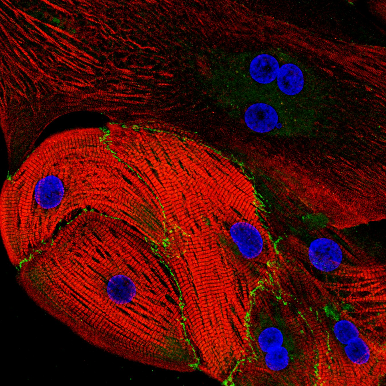
A network of human heart muscle cells (cardiomyocytes) grown from stem cells. - Blue: Cell nuclei
Green: Connections between neighboring heart muscle cells
Red: The contractile fibers that make heart muscle cells shorten and generate force
- Where is it found?
This network is located in the heart muscle, where billions of cardiomyocytes contract together to generate each heartbeat.
- How big is it?
This network measures in at about 40–70 micrometers (µm)
≈ half the width of a human hair
- What can we learn from this image?
Heart muscle cells don’t work alone. They form tightly connected networks that allow them to act as one coordinated tissue
- What are its superpowers?
Every heartbeat depends on millions of cardiomyocytes working in perfect synchrony. Their contractile fibers shorten and relax in a precisely coordinated rhythm, allowing the heart to pump blood throughout the body.
- How was this image taken?
This image was taken with a confocal microscope, which uses laser light to create sharp, highly detailed images of structures inside cells.
- Image: Eurac Research / Marzia De Bortoli – Molecular Biologist
- What are scientists trying to understand with this image?
Researchers use human cardiomyocytes from patients with inherited heart diseases to better understand what causes these conditions and to explore potential new treatments.
The story behind the image
What looks like the stripes of a zebra is actually the engine of every heartbeat. Inside each heart muscle cell, thousands of perfectly organized contractile fibers work together so that millions of cells can beat as one, keeping your heart pumping every second of your life.
Link
Credits: Eurac Research / Marzia De Bortoli