06
The hidden architecture of life
- What do you see?
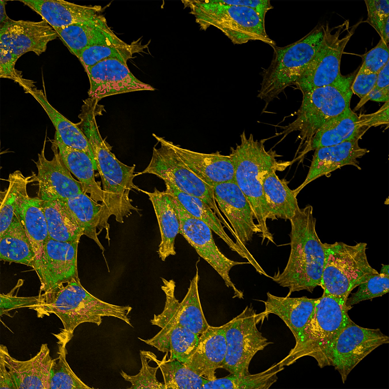
A multicolored portrait of human neuronal cells created using the Cell Painting technique. Different fluorescent dyes highlight several cellular structures at the same time, revealing the intricate organization inside each cell. - Blue: Cell nucleus
Green: Endoplasmic reticulum
Red: Mitochondria
Yellow: Several cellular structures, including the cell membrane, Golgi apparatus, and cytoskeleton
- Where are they found?
Neurons are found throughout the nervous system, including the brain and spinal cord. This image shows human neuronal cells grown in the laboratory. These cells are widely used to study how neurons function, how diseases affect them, and how they respond to potential new therapies.
- How big are they?
The entire image is approximately 100–150 micrometers (µm) wide.
≈ twice the width of a human hair
- What can we learn from this image?
A cell is far more than a tiny bag of molecules. It contains a highly organized collection of structures that each perform specialized tasks. By visualizing many of these structures at once, scientists can detect even subtle changes that may signal disease or reveal how a new treatment is working.
- What are their superpowers?
The Cell Painting technique transforms cells into a detailed biological “fingerprint”. And, by labeling multiple cellular structures simultaneously, this technique allows scientists to compare healthy and diseased cells and identify even the smallest changes caused by illness or new medicines.
- How was this picture taken?
This image was captured using a confocal microscope. Fluorescent dyes were used to label different parts of the cell, allowing the microscope to produce a detailed, high-resolution view of its internal organization.
- Image: Eurac Research / Martin Lang – Molecular Biologist
- What are scientists trying to understand with this image?
Scientists use Cell Painting to investigate how diseases alter the inner organization of cells and how potential therapies can restore healthy cellular function. Because each cell produces its own unique “fingerprint,” this approach can reveal changes that might otherwise go unnoticed.
The story behind the image
At first glance, this image looks like a colorful abstract painting. In reality, every color represents a different part of a living cell, each carrying out its own essential task. Together, these components create a unique cellular fingerprint that tells scientists how healthy the cell is, how it responds to its environment, and whether a potential new therapy is having the desired effect.
Credits: Eurac Research / Martin Lang