04
The heart is wired
- What do you see?
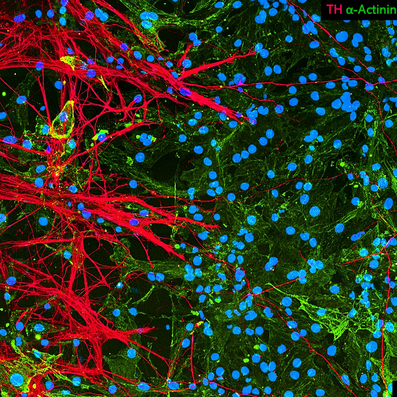
Tiny sympathetic nerve fibers (axons) communicate with heart muscle cells (cardiomyocytes), forming part of the communication network that helps regulate heartbeat. - Blue: Cell nuclei
Green: Heart muscle cells (cardiomyocytes)
Red: Sympathetic nerve fibers
- Where is it found?
In the human heart, sympathetic nerve fibers form an extensive network within the muscle.
- How big is it?
These cells measure in at about 120 micrometers (µm)
≈ 1.5 human hairs placed side by side
- What can we learn from this image?
Your heart doesn’t only beat because of its muscle cells. A dense network of nerve cells constantly fine-tunes every heartbeat, controlling how fast and how forcefully your heart beats.
- What are their superpowers?
These networks act like the heart’s accelerator. When your brain senses excitement, stress, or exercise, sympathetic nerves send signals that make the heart beat faster and contract more strongly.
- How was this image taken?
This image was taken with a confocal microscope, which uses laser light to reveal tiny biological structures in great detail
- Image: Eurac Research/ Giada Cattelan – Cellular Biologist, Alexandros Lavdas
- What are scientists trying to understand with this image?
Scientists are exploring how sympathetic neurons communicate with heart muscle cells, and how changes in this interaction may contribute to heart disease and abnormal heart rhythms (arrhythmias).
The story behind the image
Hidden within every heartbeat is an intricate communication network. Thousands of microscopic nerve fibers weave through the heart, reaching individual muscle cells like branches of a living tree. Each burst of activity can subtly change how fast and how strongly the heart beats, allowing it to respond instantly when you run, laugh, or face a challenge. Understanding this invisible wiring helps scientists uncover how the heart stays in rhythm and what happens when that rhythm is disrupted.
Link
Credits: Eurac Research / Giada Cattelan