10
Recipe for a small brain
- What do you see?
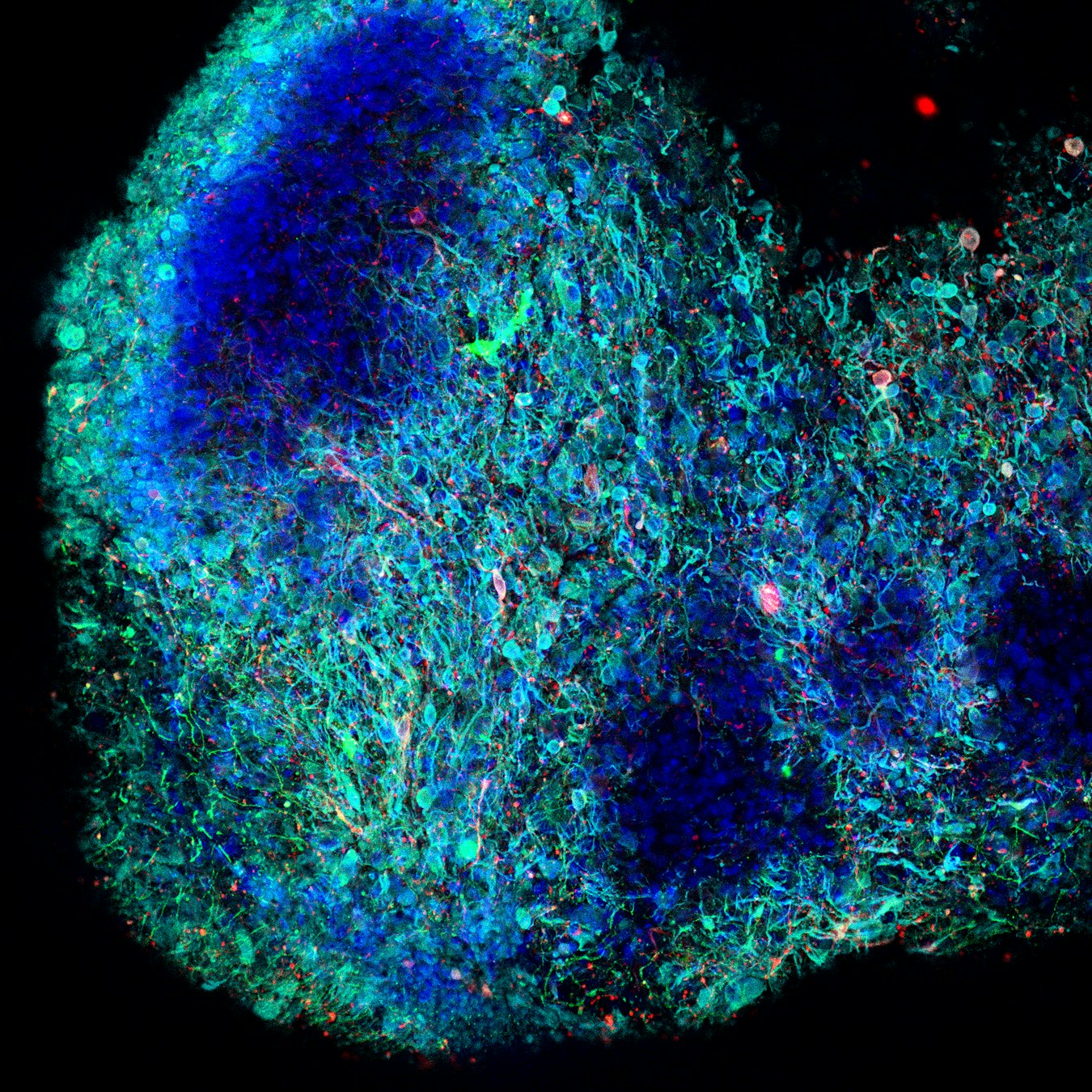
A tiny piece of brain-like tissue grown in the laboratory from adult human stem cells.
Although only about the size of a pinhead, this piece of tissue contains many of the same types of nerve cells found in the human midbrain. - Blue: The nuclei, which contain the cells’ genetic information.
Green: RIT2, a protein found in nerve cells.
Red: Cells that produce dopamine, a chemical messenger the brain uses to control movement.
Cyan: The nerve cells and their long branches.
- Where is it found?
Nowhere - at least not naturally. This is a midbrain organoid, a miniature model of part of the human brain grown in the lab to help scientists study how the brain develops and what happens when disease begins.
- How big is it?
Up to 1 millimeter in diameter.
- What can we learn from this image?
To understand how diseases such as Parkinson emerge, we have to understand which building stones lead to the disease and which ones can protect from it. Organoids are excellent three-dimensional models of human brain tissue used by researchers to experiment with different building stones, such as the protein RIT-2 in case of this picture, which depends on the genetics of the cells.
- What are its superpowers?
This tiny organoid recreates some of the brain circuits that help us move, learn, feel motivated, and experience reward.
Because the same dopamine-producing nerve cells are damaged in Parkinson’s disease, organoids like this allow scientists to study the disease in ways that would not be possible in the human brain.
- How was this image taken?
Scientists made the tissue transparent and used fluorescent labels to highlight different cell types. A powerful microscope then captured hundreds of images at different depths, which were combined into this three-dimensional view.
- Image: Eurac Research / Mattia Volta – Neuroscientist, Francesca Pischedda, Asiye Malkoç
- What are scientists trying to understand with this image?
Researchers are using organoids like this to discover how Parkinson’s disease begins. By studying cells carrying genetic changes linked to the disease, they hope to identify its earliest signs and develop better treatments.
The story behind the image
This tiny organoid is not a brain. It cannot think or feel. But by recreating some of the brain’s most vulnerable nerve cells, scientists gain a remarkable opportunity: to watch Parkinson’s Disease unfold in the laboratory and search for ways to stop it before irreversible damage occurs.
Links
Credits: Eurac Research / Mattia Volta